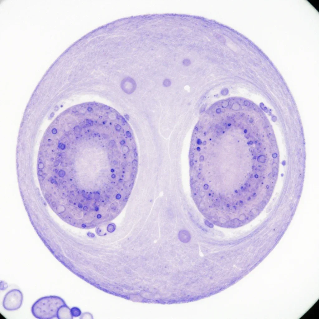
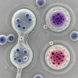
breast cells under microscope, pathology image,
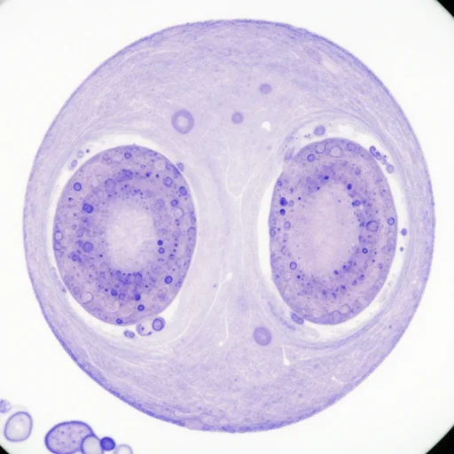
breast cells under microscope, pathology image,
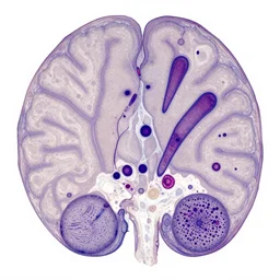
photo taken with a light microscope showing spongiform degeneration in the cerebral cortex of a patient who had died of creutzfeldt - jakob disease ( cjd ). the tissue section was embedded in paraffin - wax, sectioned at a thickness of approximately 5 - 1 0 microns, and stained with hematoxylin and eosin ( h & e ). several large, pyramid - shaped neurons can be seen, along with the nuclei of glial cells.
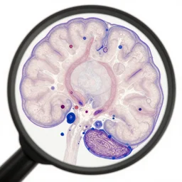
magnified 1 0 0 x, and stained with h & e ( hematoxylin and eosin ) staining technique, this light photomicrograph of brain tissue reveals the presence of prominent spongiotic changes in the cortex, and loss of neurons in a case of variant creutzfeldt - jakob disease ( vcjd ).
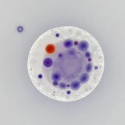
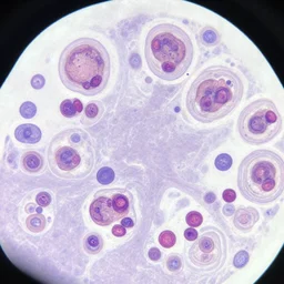
a microscopy image showing an abnormal stained nucleus
a microscopy image showing a cell with a normal nucleus and one cell with an abnormal stained nucleus, confocal miscroscopy, paper, cell journal, cell biology, nature journal, science
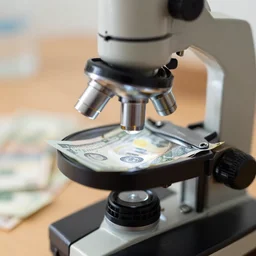
money seen under the microscope
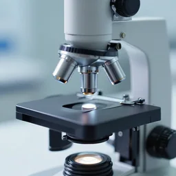
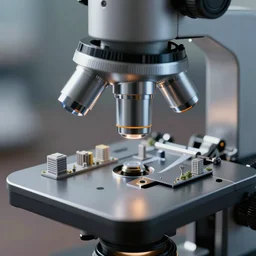
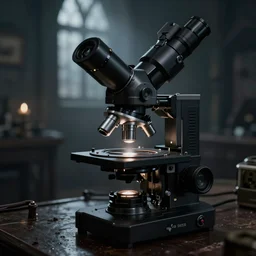
a microscopic \ ; \ n microscope
microscopic panda found in microscope
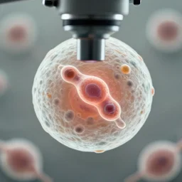
a singular cell undergoing cell division as seen through a microscope
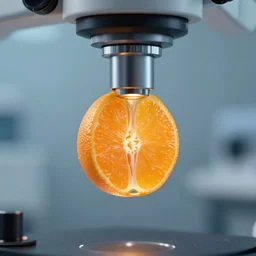
an electron microscope image of an orange segment

medical image
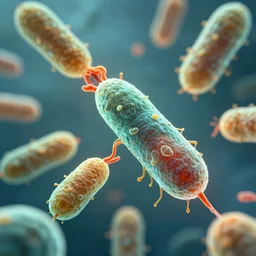
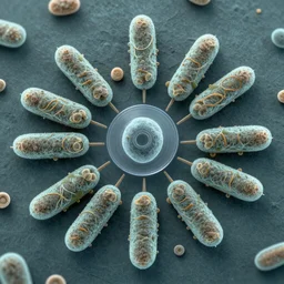
high quality render of bacteria infecting healthy cells biology virus microscope
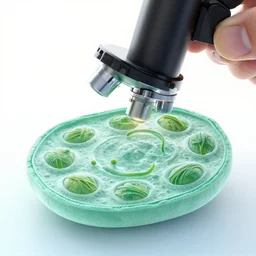
plant cell under a microscope
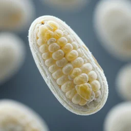
Corn under an electron microscope
microscopic city seen by microscope lens, very detailed
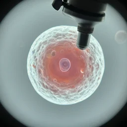
a singular living cell as seen through a microscope
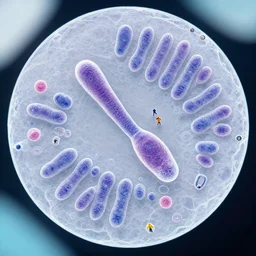
microscopy of cell in metaphase except the chromosomes are replaced by tiny humans
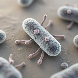
microscope photograph of a bacteria with cilia
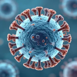
electron microscope image of a cooties virus

microscopic elvis impersonator seen in a scanning electron microscope
microscopy, 1 0 0 0 x magnification, electron microscope, diatoms, tardigrades, close up, centered, bacteria germs made in the style of roger dean
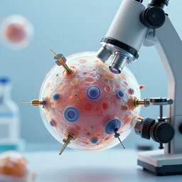
a microscope picture of an artificial cell made of machines
Electron Microscope Dark Souls

electron microscope photo of a miniscule gorilla