

a view of barcelona, scanning electron microscope style

a view of barcelona, scanning electron microscope style
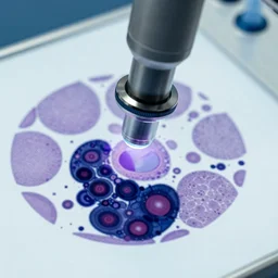
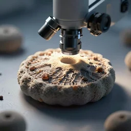
table viewed under electron microscope

microscopic view of a neuron taken with scanning tunneling electron microscope

scanning electron microscopy, highly detailed, masterpiece
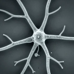
neurons and dendrites seen through scanning electron microscope

electron microscope view of a dining room table

tunneling electron microscope image of a cell pierced by an arrow. line penetrating cell. false color. mit technology review. nature journal. nobel prize winning. ultra detailed 8 k tiff

neuron cell viewed from a scanning tunneling electron microscope, ultra high detail

strands of DNA, scanning tunneling microscope, highly detailed

microscopic elvis impersonator seen in a scanning electron microscope

a very angry microbe with human eyes, scanning electron microscope

dining room table viewed through electron microscope

tunneling electron microscope image of a cell quartered by an arrow. false color. mit technology review. nature journal. nobel prize winning. ultra detailed 8 k tiff

flat lay, electron microscope knolling of the parts of a cell.
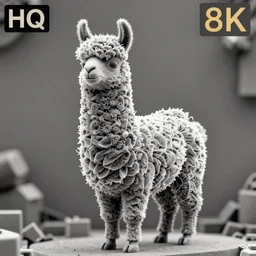
Scanning tunneling microscope image highly rhythmic expressionistic wild symmetrical nano-scale sculpture furry llama portrait in Scanning tunneling microscope, HQ 8k scan

tunneling electron microscope image of a cell pierced by a shaft of gas. line penetrating cell. false color. mit technology review. nature journal. nobel prize winning. ultra detailed 8 k tiff

a very angry microbe named jaume, scanning electron microscope

tunneling electron microscope image of a eukaryote pierced by a line of atoms. line penetrating cell. false color. mit technology review. nature journal. nobel prize winning. ultra detailed 8 k tiff

tunneling electron microscope image of a broken cell eukaryote lysed. false color. leaking cell broken membrane. mit technology review. nature journal. nobel prize winning. ultra detailed 8 k tiff
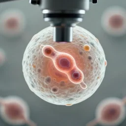
a singular cell undergoing cell division as seen through a microscope

tunneling electron microscope image of a mitochondria organelle. false color. mit technology review. nature journal cover. nobel prize winning. ultra detailed 8 k tiff

tunneling electron microscope image of oxygen migrating across the cell membrane. penetrating crossing cell membrane edge. dividing membrane. false color. mit technology review. cell journal. nobel prize winning. ultra detailed 8 k tiff
a diatom viewed through an electron microscope.

scanning electron micrograph of a human t cell