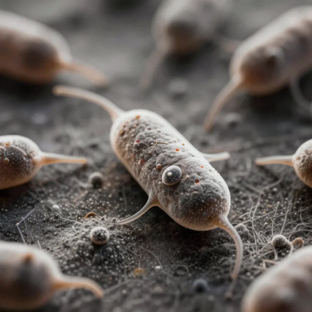

diagram of eukaryotic cell with labels
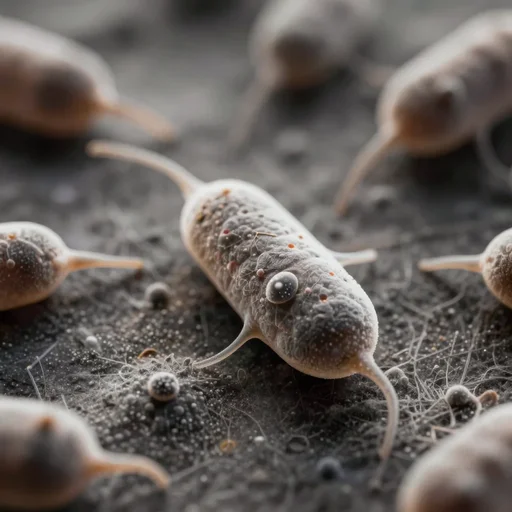
diagram of eukaryotic cell with labels

a black and white close - up painting of a single!! eukaryotic cell showing the nucleus and mitochondria

a black and white detailed anatomy of a single!! eukaryotic cell showing the nucleus and mitochondria

a black and white detailed anatomy of a single! eukaryotic cell showing the nucleus, mitochondria, golgi and endoplasmic reticulum

tunneling electron microscope image of leaking cell membrane, broken leaking cell wall, liquid oxygen migrating across cell membrane of a mitochondria oblong organelle. liquid stream penetrating crossing cell membrane edge. dividing crossing over eukaryote membrane. false color. mit technology review. cell science nature. nobel prize winning. ultra detailed 8 k tiff

an illustration of the inside of an eukaryotic cell by picasso, cubic period

a black and white detailed anatomy of a single!! eukaryotic cell showing the nucleus and mitochondria, engraved on wood

tunneling electron microscope image of broken membrane, broken leaking cell wall, liquid oxygen migrating across the cell membrane of a mitochondria oblong organelle. liquid stream penetrating crossing cell membrane edge. dividing crossing over eukaryote membrane. false color. mit technology review. cell science nature. nobel prize winning. ultra detailed 8 k tiff

universe star field flowing into single cell nucleus eukaryote molecular

tunneling electron microscope image of broken membrane, broken leaking cell wall, liquid oxygen migrating across the cell membrane of a mitochondria oblong organelle. liquid dots penetrating crossing cell membrane edge. dividing crossing over eukaryote membrane. false color. mit technology review. cell journal. nobel prize winning. ultra detailed 8 k tiff

a biology textbook illustration of an eukaryotic cell, surrealistic, black and white

a bacterial cell

tunneling electron microscope image of liquid oxygen migrating across the cell membrane of a mitochondria oblong organelle. liquid dots penetrating crossing cell membrane edge. dividing crossing over eukaryote membrane. false color. mit technology review. cell journal. nobel prize winning. ultra detailed 8 k tiff

tunneling electron microscope image of broken membrane, broken leaking cell wall, liquid oxygen migrating across the cell membrane of a mitochondria oblong organelle. liquid dots penetrating crossing cell membrane edge. dividing crossing over eukaryote membrane. false color. mit technology review. cell science nature. nobel prize winning. ultra detailed 8 k tiff

a biology textbook illustration of eukaryotic cells, surrealistic, black and white
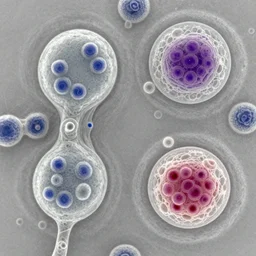
a microscopy image showing a cell with a normal nucleus and one cell with an abnormal stained nucleus, confocal miscroscopy, paper, cell journal, cell biology, nature journal, science

tunneling electron microscope image of a broken cell eukaryote lysed. false color. leaking cell broken membrane. mit technology review. nature journal. nobel prize winning. ultra detailed 8 k tiff

a black and white detailed anatomy of an eukaryotic cell showing the mucleus, mitochondria, golgi and endoplasmic reticulum, painting by da vinci

a biology textbook illustration of eukaryotic cells, cubic style, black and white

in jail cell

a black and white detailed anatomy of an eukaryotic cell showing the mucleus, mitochondria, golgi and endoplasmic reticulum, painting by mc escher
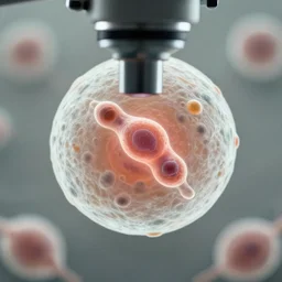
a singular cell undergoing cell division as seen through a microscope

tunneling electron microscope image of a eukaryote pierced by a line of atoms. line penetrating cell. false color. mit technology review. nature journal. nobel prize winning. ultra detailed 8 k tiff

modern norway prison cell